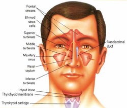
Ethmoidal sinuses
These multiple, thin-walled cavities (cells) occupy the whole of the ethmoidal labyrinth, between the orbit and the upper part of the cavity of the nose. They form three groups, of which the anterior and middle cells open into the superior meatus. Explore them with a blunt probe, and attempt to find their opening into the nasal cavity.
Orbita periosteum
The orbital periosteum form a funnel-shaped sheath which is loosely attached to the bony walls, and encloses all the contents of the orbit except the zygomatic nerve and the infra-orbital nerve and vessels. It is continuous with the endocranium through the optic canal and the superior orbital fissure.
Rectus superior
It arises from the upper margin of the optic canal,passes anterolaterally above the optic nerve, and is inserted into the sclera about 6mm posterior to the sclerocorneal junction. Nerve supply: superior division of the oculomotor nerve.
Obliquus superior
It arises from the roof of the orbit immediately anteromedial to the optic canal, and passes anteriorly along the upper part of the medical orbital wall. Anteriorly, it end in a slender tendon which enters the trochlea, and at once turns posterolaterally to pass between the superior rectus and the eyeball. Lateral to the superior rectus, the tendon flattens out and inserted into the sclera midway between the entrance of the optic nerve and the cornea. Nerve supply: the trochlear nerve. Action: it turns the cornea downwards when it is already turned medially; a position in which the inferior rectus is ineffective as a depressor of the cornea. The trochlea is a small fibrocartilaginous ring attached by fibrous tissue to the trochlear fossa on the frontal bone. It is lined with asynovial sheath which allows the tendon to slide freely in it.
