Sutural ligaments
The foosely attached pericranium is continuous with the endocranium through the suture of the skull, forming the sutural ligaments. These hold the bones together and allow growth between them. As the skull consolidates from the third to fourth decade onwards, the adjacent bones unite by ossification of the sutural ligaments-a process which begins internally and is known as synostosis.
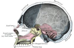
Endocranium
When the skull cap is detached, the outer surface of the endocranium is exposed. It is rough because of the fine fibrous and vascular processes which pass between it and the bones. Torn blood vessels are most numerous close to the midline. Here one of the largest intracranial venous channels lies deep to the endocranium. If a blunt instrument is pressed on the sinus, blood oozes from the numerous, small veins which have been ruptured. The endocranium is more firmly attached to the base of the cranial cavity than to the vault, the degree of adhesion varying with age and from individual to individual.
A number of branching vessels ascend on the outer surface of the endocranium towards the vertex. These branches of the middle meningeal artery, with the corresponding veins on their external surfaces, groove the inner table of the skull, and out in relief from the surface of the endocranium. They supply the skull the endocranium, and the dura mater which is fused to its internal surface. These meningeal vessels play on part in supplying the pia-arachnoid or the brain itself, but may be torn in fractures of the skull. This results in arterial bleeding between the endocranium and the skull (extradural haemorrhage) rapidly producing a swelling which presses on the brain. If the fracture tears the endocranium and dura mater (the outermost of the meninges), then bleeding may spread into the space (subdural space) which separates the dura from the other meninges covering the brain-a subdural haemorrhage.
A number of branching vessels ascend on the outer surface of the endocranium towards the vertex. These branches of the middle meningeal artery, with the corresponding veins on their external surfaces, groove the inner table of the skull, and out in relief from the surface of the endocranium. They supply the skull the endocranium, and the dura mater which is fused to its internal surface. These meningeal vessels play on part in supplying the pia-arachnoid or the brain itself, but may be torn in fractures of the skull. This results in arterial bleeding between the endocranium and the skull (extradural haemorrhage) rapidly producing a swelling which presses on the brain. If the fracture tears the endocranium and dura mater (the outermost of the meninges), then bleeding may spread into the space (subdural space) which separates the dura from the other meninges covering the brain-a subdural haemorrhage.
