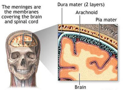
Dura mater
This has been seen and described during the dissection of the head but its parts should bereviewed in relation to the brain.
Arachnoid
This exceedingly thin, almost transparent membrane lines the internal surface of the dura mater, and has exactly the same shape as the dural sac except where its arachnoid granulations pierce the dura mater. The arachnoid is separated from the dura mater by a bursa-like, capillary space containing a film of fluid. This forms a sliding plane where movement is possible between the dura mater and the brain enclosed in the arachnoid and pia mater, except where the arachnoid and dura are fused, i.e where both are pierced by structures entering or leaving the brain (e.g. nerves and blood vessels), where the arachnoid granulations pierce the dura mater, and where the ligamenta denticulata are attached to the dura mater.
Sub arachnoid space
This space, between the arachnoid and pia mater is filled with cerebrospinal fluid which enters it from the cavities (ventricles) of the brain, and acts as a mobile buffer to distribute and pressureswithin the skull.
Filaments or trabeculae traverse the subarachnoid space from the arachnoid to the pia mater, and arenumerous in some situations, e.g. on the surfaces of the cerebral hemispheres. Here the dense trabeculae form a kind of fluid-filled sponge which may to protect the surface of the brain from damage against the skull and the dural folds (the falx and tentorioum) when it moves withinthe dura mater. They also bind the pia and arachnoid tightly together in these situations. Elsewhere, and especially where the pia mater, closely investing the brain, is widely separated from the arachnoid lining the dura mater, the mesh is less dense and the cerebrospinal fluid can flow more freely. The large arteries and veins of the brain lie in the subarachnoid space, and entering or leaving the brain traverse it. In some situation, e.g. the stem of the lateral sulcus, the space forms a tight sleeve around the artery, so that its pulsations force the cerebrospinal fluid along the sleeve. Where the vessels on the external surface of the pia mater send branches into the substance of the brain, a sleeve of pia mater and subarachnoid space (perivascular space) surrounds each branch for a short distance until the pia mater fuses with the wall of vessel.
Filaments or trabeculae traverse the subarachnoid space from the arachnoid to the pia mater, and arenumerous in some situations, e.g. on the surfaces of the cerebral hemispheres. Here the dense trabeculae form a kind of fluid-filled sponge which may to protect the surface of the brain from damage against the skull and the dural folds (the falx and tentorioum) when it moves withinthe dura mater. They also bind the pia and arachnoid tightly together in these situations. Elsewhere, and especially where the pia mater, closely investing the brain, is widely separated from the arachnoid lining the dura mater, the mesh is less dense and the cerebrospinal fluid can flow more freely. The large arteries and veins of the brain lie in the subarachnoid space, and entering or leaving the brain traverse it. In some situation, e.g. the stem of the lateral sulcus, the space forms a tight sleeve around the artery, so that its pulsations force the cerebrospinal fluid along the sleeve. Where the vessels on the external surface of the pia mater send branches into the substance of the brain, a sleeve of pia mater and subarachnoid space (perivascular space) surrounds each branch for a short distance until the pia mater fuses with the wall of vessel.
